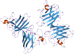
Superoxide dismutase

Order Now!
Superoxide Dismutase (SOD) is also known as Orgotein, SOD, Super Dioxide Dismutase, Superóxido Dismutasa, Superoxydase Dismutase, or Superoxyde Dismutase. The SOD1 gene is located on the long (q) arm of chromosome 21 at position 22.11. More precisely, the SOD1 gene is located from base pair 33,031,934 to base pair 33,041,243 on chromosome 21.
Superoxide dismutase is a metalloenzyme. It is present in all living organisms, including animals, plants, and microorganisms. SOD is categorized based on its metallicity into copper, zinc-SOD (Cu, Zn-SOD), manganese-SOD (Mn-SOD), and iron-SOD (Fe-SOD). LifeTein's recombinant Superoxide Dismutase belongs to Human Copper, Zinc-Superoxide Dismutase (rh-SOD1).
LifeTein's Superoxide Dismutase1 (SOD1) is expressed and purified from E.coli. The N terminal sequence is ATKAVCVLKG. This enzyme binds to molecules of copper and zinc to break down toxic, charged oxygen molecules called superoxide radicals. The physiological significance of SOD is that it can convert toxic superoxide free radicals into hydrogen peroxide.
Superoxide radicals can damage cells if too many accumulate within cells. Superoxide radicals are byproducts of normal cell processes, particularly energy-producing reactions, and must be broken down regularly. It is living organisms’ primary means of scavenging oxygen free radicals.
Almost 60 diseases have been shown to be directly related to oxygen free radicals and the level of SOD has used as an indicator of aging and death. SOD has recently gained notoriety for its connection with amyotrophic lateral sclerosis (ALS), more commonly known as Lou Gehrig's disease, a condition characterized by progressive movement problems and muscle wasting. This disease is a degenerative disorder that leads to selective death of neurons in the brain and spinal cord, leading to gradually increasing paralysis. At least 170 mutations in the SOD1 gene have been found to cause ALS. Most of these mutations change one of the amino acids in the superoxide dismutase enzyme. About half of all Americans with ALS caused by SOD1 gene mutations have a particular mutation that replaces the amino acid alanine with the amino acid valine at position 4 in the enzyme.
It is unclear why the nerve cells are particularly sensitive to SOD1 gene mutations. Researchers have suggested that the altered enzyme may cause the death of nerve cells. These possibilities include an accumulation of harmful superoxide radicals, increased production of other types of toxic radicals, increased cell death, or formation of aggregates of misfolded superoxide dismutase that may be toxic to cells.
Studies have shown that SOD can play a critical role in reducing internal inflammation and lessening pain associated with conditions such as arthritis. Supplement with oral preparations of pure SOD enzyme is not effective because the SOD protein is easily deactivated by acids and enzymes contained in the digestive tract. However scientists have created bioavailable forms of SOD using natural plant extracts. When delicate SOD molecules are coupled with a protective protein derived from wheat and other plants, they can be delivered intact to the intestines and absorbed into the bloodstream, thus effectively enhancing the body’s own primary defense system.
SOD is a key component in cosmetic products and has been approved by many countries because it can delay aging, regulate immune response, and blood lipid levels, and prevent damage from solar radiation. SOD is an unusually stable enzyme although its apoenzyme is very unstable.
Full sequence |
MATKAVCVLK GDGPVQGIIN FEQKESNGPV KVWGSIKGLT EGLHGFHVHE FGDNTAGCTS
AGPHFNPLSR KHGGPKDEER HVGDLGNVTA DKDGVADVSI EDSVISLSGD HCIIGRTLVV
HEKADDLGKG GNEESTKTGN AGSRLACGVI GIAQ |
Function |
Destroys radicals which are normally produced within the cells and which are toxic to biological systems. |
Catalytic activity |
2 superoxide + 2 H+ = O2 + H2O2. |
Cofactor |
Binds 1 copper ion per subunit.
Binds 1 zinc ion per subunit. |
Subunit structure |
Homodimer; non-disulfide linked. Homodimerization may take place via the ditryptophan cross-link at Trp-33. The pathogenic variants ALS1 Arg-38, Arg-47, Arg-86 and Ala-94 interact with RNF19A, whereas wild-type protein does not. The pathogenic variants ALS1 Arg-86 and Ala-94 interact with MARCH5, whereas wild-type protein does not. |
Subcellular location |
Cytoplasm. Note: The pathogenic variants ALS1 Arg-86 and Ala-94 gradually aggregates and accumulates in mitochondria. |
Post-translational modification |
Unlike wild-type protein, the pathogenic variants ALS1 Arg-38, Arg-47, Arg-86 and Ala-94 are polyubiquitinated by RNF19A leading to their proteasomal degradation. The pathogenic variants ALS1 Arg-86 and Ala-94 are ubiquitinated by MARCH5 leading to their proteasomal degradation.
The ditryptophan cross-link at Trp-33 is responsible for the non-disulfide-linked homodimerization. Such modification might only occur in extreme conditions and additional experimental evidence is required. |
Involvement in disease |
Amyotrophic lateral sclerosis 1 (ALS1): A neurodegenerative disorder affecting upper motor neurons in the brain and lower motor neurons in the brain stem and spinal cord, resulting in fatal paralysis. Sensory abnormalities are absent. The pathologic hallmarks of the disease include pallor of the corticospinal tract due to loss of motor neurons, presence of ubiquitin-positive inclusions within surviving motor neurons, and deposition of pathologic aggregates. The etiology of amyotrophic lateral sclerosis is likely to be multifactorial, involving both genetic and environmental factors. The disease is inherited in 5-10% of the cases.
Note: The disease is caused by mutations affecting the gene represented in this entry. |
Miscellaneous |
The protein (both wild-type and ALS1 variants) has a tendency to form fibrillar aggregates in the absence of the intramolecular disulfide bond or of bound zinc ions. These aggregates may have cytotoxic effects. Zinc binding promotes dimerization and stabilizes the native form. |
Sequence similarities |
Belongs to the Cu-Zn superoxide dismutase family. |
Additional reading on superoxide dismutase:
P. Pasinelli and R. H. Brown (2006) Molecular biology of amyotrophic lateral sclerosis: insights from genetics. Nature Reviews Neuroscience 7, 710-721.
J. A. Imlay and I. Fridovich (1991) Assay of metabolic superoxide production in Escherichia coli. Journal of Biological Chemistry 266, 6957-6965.
I. Fridovich (1989) Superoxide dismutases. Journal of Biological Chemistry 264, 7761- 7764.
E. D. Getzoff, J. A. Tainer, P. K. Weiner, P. A. Kollman, J. S. Richardson and D. C. Richardson (1983) Electrostatic recognition between superoxide and copper, zinc superoxide dismutase. Nature 306, 287-290.